Neuroscience. 2015 Aug 20;301:90-105. doi: 10.1016/j.neuroscience.2015.05.062.
Pathological changes in hippocampal neuronal circuits underlie age-associated neurodegeneration and memory loss: positive clue toward SAD.
Moorthi P, Premkumar P, Priyanka R, Jayachandran KS, Anusuyadevi M
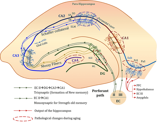
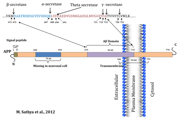
Figure 1. Anatomical view of rat hippocampus: Schematic representation of hippocampal circuits showing, segments of Cornu ammonis (CA) in hippocampal formation (Layers of the pyramidal neurons: Stratum Oriens (SO), Stratum Pyramidale (SP), Stratum Radiatum (SR), Stratum lacunosum moleculare (SLM), Stratum Lucidum (SL)), Dentate gyrus (DG) (Three layers: Molecular Layer (ML), Granule Cell Layer (GCL) & Subgranular zone (SGZ)) and subiculum). Subiculum is the most inferior component of the hippocampal formation (Tsien et al., 2013; Llorens-Martin et al., 2014). It lies between the CA1 subfield of hippocampus proper and entorhinal cortex. It receives information from CA1 (glutamatergic transmission) and entorhinal cortical layer III pyramidal neurons and is the main output of the hippocampus (Kintscher et al., 2012). The Hippocampus proper is a composed series of Cornu Ammonis areas: first CA4 (which underlies the DG), then CA3, then a very small zone called CA2, and then CA1. Hippocampal proper is filled with densely packed pyramidal cells. The mossy fibers terminate in a relatively narrow zone mainly located just above the CA3 pyramidal cell layer. The dentate projection to CA3 stops near the border of CA3 and CA2, and the lack of granule cell input is one of the main features that distinguish CA3 from CA2 pyramidal cells (Insausti and Amaral, 2004). While, DG have three layers, there is a relatively cell-free layer called the molecular layer (ML) which is occupied by the dendrites of the dentate granule cells and fibers of the perforant path that originate from entorhinal cortex (EC). Arrow heads indicating neuronal transmission that connect different neuronal circuits of trisynaptic circuit (ECII-DG-CA3-CA1), monosynaptic circuit (ECII-CA1), perforant pathway, which are involved in the consolidation and reconsolidation of the memory. Schematic representation showing trisynaptic and monosynaptic connections & highlights the age-associated sequential pathological changes in hippocampal circuits connecting various subregions (CA1, CA2, CA3, DG and subiculum). Damages in these neuronal circuits may a possible cause of age associated memory loss.
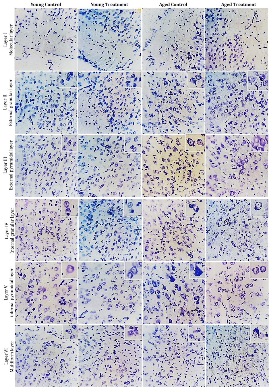
Figure 2. CV staining of a section obtained from the neocortex: An illustrative slice of the neocortex, in comparison to typical six-layered normal neocortex (uppercase Roman numerals). Cortical layers could be easily distinguished by cell size, shape, and density. B) Schematic representation of neocortex showing six-layered architecture: Layer I (Molecular layer) consists of very few neurons, contains horizontal cell axons and dendrites from pyramidal cells (predominantly glutamatergic and GABAergic but also include cholinergic, noradrenergic, and serotoninergic axons) in other layers of the Neocortex; Layer II (External Granular layer) consists of granule cells, whose dendrites reach to the molecular layer and axons to deeper layers; Layer III (External pyramidal layer) consists of two sub layers of pyramidal cells, a superficial layer of medium cells and a deeper layer of large cells, Their dendrites reach to layer I, and axons to other cortical areas (Chu et al., 2003; Petersen and Crochet, 2013). The superficial layer to areas on the same side and the deeper layer to areas in the other hemisphere; Layer IV (Internal Granular layer) consists of stellate cells, which mediate between inputs from other areas to pyramidal dendrites and axons, and from pyramidal dendrites and axons (Walcott and Langdon, 2002; Petersen and Crochet, 2013). Often divided into two sublayers IVa and IVb; Layer V (Internal Pyramidal layer) consists of medium and large sized pyramidal cells intermingled with granule and Martinotti cells, The dendrites of the large-sized pyramidal cells reach to layer I, the dendrites of the small-sized pyramidal cells reach only to layer IV, or stay within layer V; Layer VI (Multiform layer) consists of spindle cells with axons perpendicular to the cortical surface. Larger ones send dendrites to layer I, and smaller ones to layer IV (Hedreen et al., 1991; Andjelic et al., 2009).
|
Parietal cortex
|
Temporal cortex
|
Figure 3. Photomicrograph of CV stained coronal section of Parietal and Temporal cortex. A) Low magnification (10X) of parietal cortex showing six layers; B) High-magnification (40X) images of Layer I (Molecular layer, ML), Layer II (External granular layer, EGL) and Layer III (External pyramidal layer, EPL); C) Layer IV (Internal granular layer, IGL), Layer V (Internal pyramidal layer, IPL) and Layer VI (Multiform layer, MFL). Inside box shows the individual neurons, intact neuron (arrow) and dead cells (arrowhead).
Amyloid precursor protein cleavage patterns